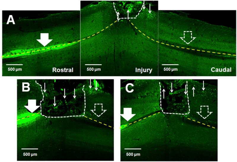
Figure 3. Crym:GFP corticospinal tract (CST) labeling 10 weeks post-SCI and bridge implantation.
In all panels, photomicrographs are from the horizontal plane, the region rostral to the lesion is to the left, the region caudal to the lesion is to the right, and the lesion site is oriented so as to be at the top. White dashed lines indicate the bridge-tissue interface. Yellow dashed lines indicate the midline of the spinal cord. (A-C) Crym:GFP expression was predominantly apparent in the dorsal column CST rostral (large solid white arrows) to the site of bridge implantation, as well as in fibers within, and exiting, the bridge (small white arrows). Minimal or no Crym:GFP expression was observed in the dorsal columns caudal to the site of bridge implantation (large open white arrows). Note that numerous Crym:GFP fibers exiting the bridge were located at the lateral edge of the horizontal section, and were not adjacent to potential regions of caudal dorsal column sparing. A, B and C each illustrate different animals, with the CST in different locations relative to the epicenter depending on section selection.

