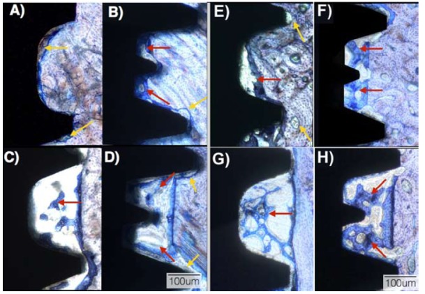
Figure 2.

Optical micrographs at 2 weeks in vivo for the (a) SW recommended instrumentation, (b) Unitite recommended instrumentation, (c) SW overdrilling instrumentation, and (d) Unitite overdrilling instrumentation. Optical micrographs at 4 weeks in vivo for the (e) SW recommended instrumentation, (f) Unitite recommended instrumentation, (g) SW overdrilling instrumentation, and (h) Unitite overdrilling instrumentation. The red arrows depict newly formed bone at the healing chambers regions; yellow arrows depict bone remodeling regions.
