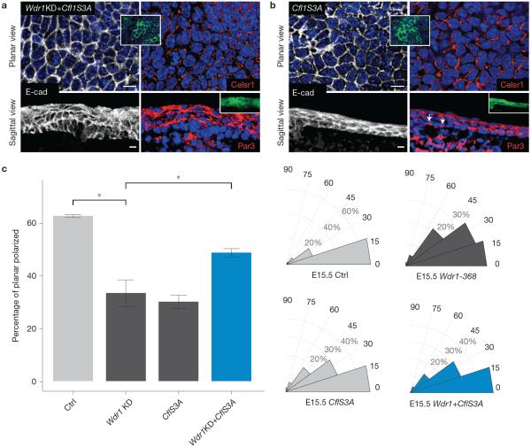
Figure 4.
Rescue of the planar polarity defects of Wdr1 mutants by expression of constitutively active cofilin. (a,b) Whole-mount immunofluorescence of E15.5 backskins co-labelled for E-cadherin and Celsr1 or sagittal views labelled with E-cadherin and Par3. (a) Wdr1-368+Cfl1S3A overexpression. (b) Cfl1S3A overexpression. (c) Quantification of data shown in a,b. For the percentage of planar-polarized cells, P = 0.00142 (Wdr1 versus Ctrl), 0.00207 (Cfl1S3A versus Ctrl), 0.247 (Wdr1+Cfl1S3A versus Ctrl), 0.0479 (Wdr1 versus Wdr1+CflS3A), ANOVA followed by Tukey's HSD test, n = 3 embryos per condition. For histograms, n = 91 cells (Ctrl); n = 82 cells (Wdr1-368); n = 131 cells (Cfl1S3A); n = 176 cells (Wdr1KD+Cfl1S3A). Error bars indicate mean ± s.e.m. Asterisks indicate statistical significance at P < 0.05. Scale bars, 10 μm.

