
Fig 6.
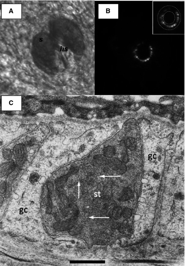
Lanceolate endings stain with FM1-43. (A) Electron micrograph through a lanceolate ending. (A) Brightfield image of a hair follicle in mouse ear skin in situ, from the dermal aspect showing the sebaceous gland (s) surrounding a central hair shaft (hs). Scale bar (also for B) indicates 50 μm. (B) FM1-43 fluorescence of lanceolate ending in the same follicle as (A), showing the appearance of the endings viewed along their length, orthogonal to their long axis. Note the almost complete circle of lanceolate terminals, forming a palisade to effectively detect hair movement in almost any direction. The inset shows the area of analysis used to measure follicle-ending fluorescence intensity. The intensity of a nearby background area was then subtracted to determine the final net intensity. (C) The more darkly stained sensory terminal (st) can be seen to be almost completely surrounded by glial cell processes (gc). SLVs present in the terminal axoplasm are indicated by white arrows. Scale bar: 0.5 μm. Micrograph from Banks et al. (2013).