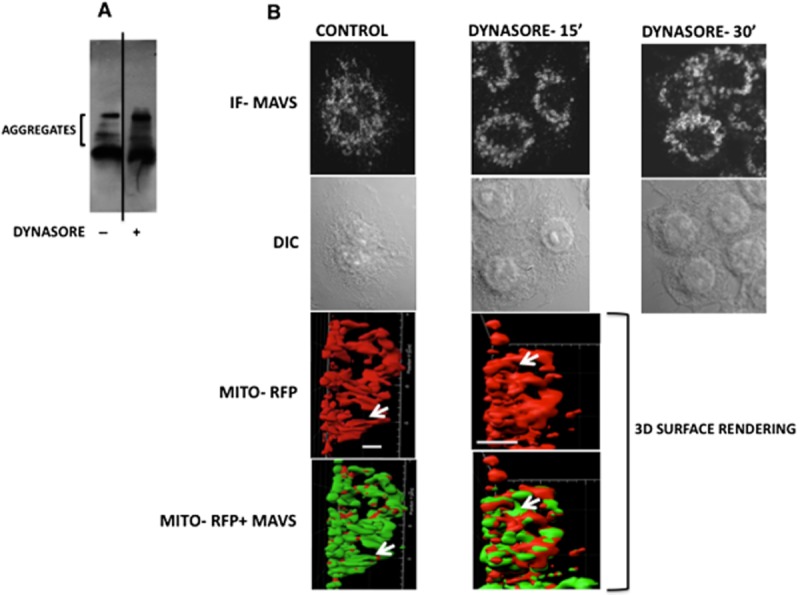
Figure 5.

Dynasore treatment causes formation of high molecular weight aggregates of MAVS. (A) SDD-AGE: RAW cells were treated with dynasore (80 μM) or DMSO for 15 min in HBSS. Cell lysates were run on SDD-AGE and immunoblotted with MAVS antibody. (B) Immunofluorescence: cells were treated with dynasore (80 μM) or DMSO for 15 and 30 min in HBSS. Cells were then fixed and subjected to fluorescent confocal microscopy (IF-MAVS) or differential interference contrast microscopy (DIC). Note the disruption of elongated MAVS thread-like structure following treatment with dynasore. Lower two panels: three-dimensional surface rendering colocalization of MAVS (immunofluorescent, green) and mitochondria (Mito-RFP, red) and changes in their structures following treatment with dynasore. Cells were transfected with Mito-RFP plasmid and treated with DMSO or dynasore (80 μM) for 15 min. Images depict side view of micrographs shown in Supporting Information Fig. S4 from Videos S1 and S2 3D surface rendering representing one frame each from control and dynasore-treated cells. Note that in control cells, MAVS covers the majority of the mitochondrial surface area indicating its even distribution on the mitochondrial surface. In dynasore-treated cells, MAVS is redistributed in a polar fashion between the mitochondria and facilitate attachment of individual threads to form multi-organelle structure of mitochondria. Bars represent 5 μm.
