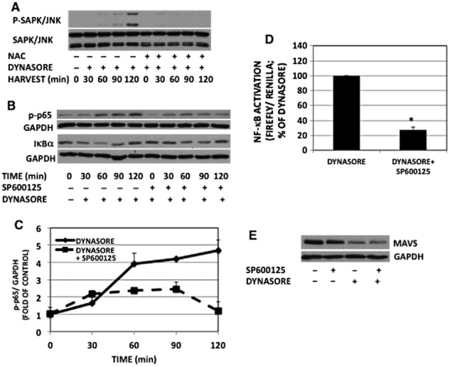
Figure 9.
Dynasore effect on NF-κB- p65 activation is mediated by SAPK/JNK. (A) Western immunoblot of phospho-SAPK/JNK: RAW cells were pretreated with or without NAC for 30 min, followed by incubation with dynasore (80 μM) for the times indicated in 2% FBS. Note the inhibition of dynasore-induced SAPK/JNK phosphorylation by NAC. (B) Western immunoblot of NF-κB-p-p65 and IκBα: cells were pre-incubated for 30 min with the SAPK/JNK inhibitor SP600125 (10 μM) or DMSO in HBSS, then dynasore (80 μM) was added to the cells for the times indicated. NF-κB-p-p65 and IκBα representative blots were from two independent separate experiments. Therefore, loading was ascertained by reprobing each corresponding blot with GAPDH. (C) Densitometry of blots of NF-κB-p-p65 and GAPDH depicted in panel B: NF-κB-p-p65 levels were normalized with GAPDH corresponding bands. (anova: P < 2 × 10⁻⁵; d.f. within groups = 20). (D) NF-κB-luciferase: SP600125 pretreatment of cells inhibited dynasore-induced enhancement of NF-κB-luciferase (P < 3 × 10⁻⁵; n = 3; Student’s t-test). (E) Western immunoblot of MAVS: cells were pre-incubated for 30 min with SP600125 (10 μM) or DMSO in HBSS, then dynasore (80 μM) or DMSO was added to the cells for an additional 15 min. Cell extracts were subjected to Western blot with anti-MAVS and GAPDH antibodies.

