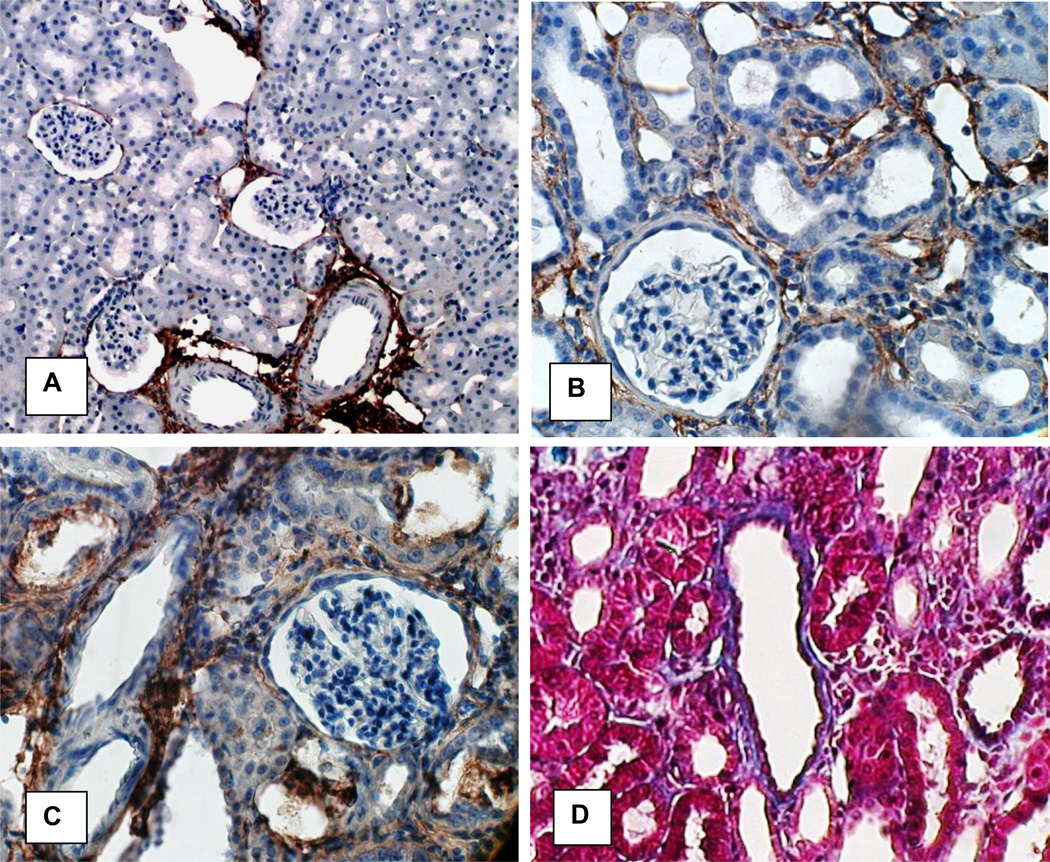
Figure 11.
Immunohistochemical analyses of the kidneys of control and HLP-fed rats for collagen. A In normal Collagen 1 expression is mostly limited to areas around large and small blood vessels. Original Mag ×10. B. C. In hyperoxaluric kidneys Collagen 1 expression is interstitial and more intense around tubules with CaOx crysral deposits. Crystals have been lost during the processing. D. Collagen stained blue with Mason’s trichrome. There is strong staining around the tubules with CaOx crystal deposits. Epithelial cells of the tubules without crystals stained red. Original Mag ×45.

