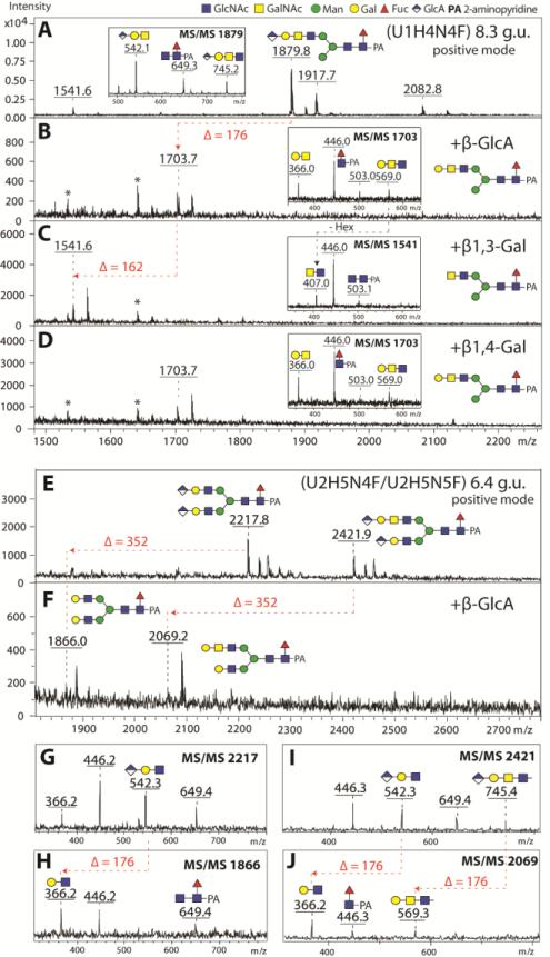
Figure 9. Structure elucidation of the glucuronylated N-glycans.
Positive ion mode MALDI-TOF/TOF MS spectra of selected RP-Amide HPLC fractions (8.3 and 6.4 g.u.) of Aedes PNGase F released anionic N-glycans before (A, E) and after treatment with β-glucuronidase (β-GlcA; B, F) and subsequent β1,3-specific (C) or β1,4-specific (D) galactosidases. Successful removal of glucuronic acid and β1,3-linked galactose moieties are indicated by the loss of 176 and 162 Da respectively. Removal of two GlcA residues from the biantennary glucuronylated N-glycan structures (m/z 2217 and 2422) is indicated by the loss of 352 Da (F). Insets in the spectra represent positive ion mode MS/MS spectra of untreated parent (m/z 1879) or exoglycosidase product glycan species (m/z 1703, 1541 and 1703). Panels G-J show the MS/MS for the parent and digested forms of the diglucuronylated glycans whose MS spectra are shown in E and F. Asterisks are indicating contaminating signals from the enzyme preparations. The proposed structure of the major glycan in each spectrum is also shown.

