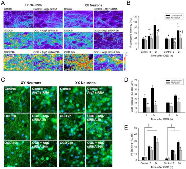
Figure 5. Oxygen-glucose deprivation (OGD) produces sex-dependent autophagy in primary cortical neurons from GFP-LC3+/− mice.
The GFP signal was enhanced immunohistochemically. A. Identification of GFP-LC3 enriched vesicles consistent with autophagosome formation at 2 and 24 h after OGD. B. Quantification of GFP-LC3 fluorescent intensity peaks over threshold (fluorescence in control cells) showing that Atg7 siRNA reduces OGD-induced autophagosome formation in XY- and XX-neurons (n = 6/group; *P < 0.05 vs. control siRNA, †P < 0.05 vs. control; ANOVA with Tukey's test). C. GFP immunohistochemistry showing neuronal loss and formation of GFP-enriched vesicles after OGD. D and E. Treatment with Atg7 siRNA reduced LDH release and increased neuron survival detected by PI labeling at 2 and 24 h after OGD in both XY- and XX-neurons vs. control siRNA (LDH, n = 6/group; PI, n = 4/group; *P < 0.05 vs. control siRNA; ANOVA with Tukey's test).

