
Figure 1. Surface and schematic view of the interaction between murine IRE1α and MKC9989 adapted from Sanches et al. [73].
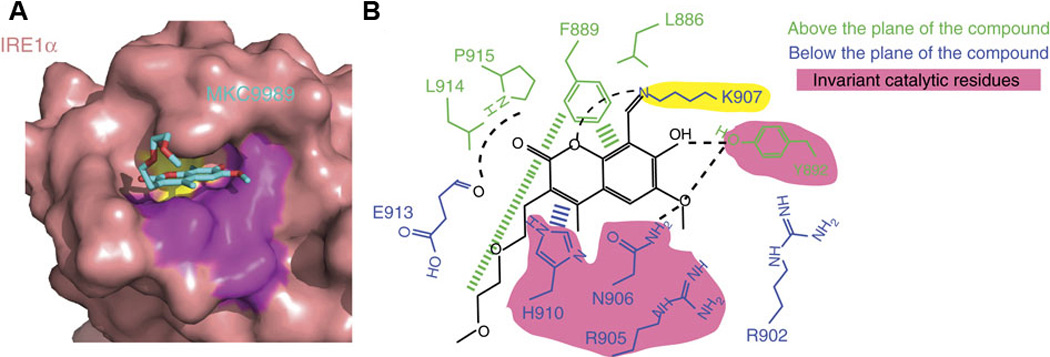
A. Surface view of the IRE1α–MKC9989 complex. Lysine 907 is colored yellow and invariant active site residues are colored purple. B. Schematic view of the contact residues of IRE1α and notable interactions between IRE1α and MKC9989.