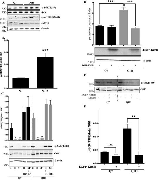
Figure 4.
mTORC1 basal activity is increased in STHdhQ111 cells. A. Western blot analysis of total p70S6K (S6K), phospho-S6K(T389), phospho-mTOR(S2448) and total mTOR expression in three independent STHdhQ7 and Q111 cell lysates. β-actin was used as the loading control. B. mTORC1 activation as quantified by the ratio of phospho-p70S6K to total p70S6K in STHdhQ7 and Q111 cells (***P < 0.001, student's t-test, n=3). C. Changes of mTORC1 activity during nutrient starvation and recovery. Cells were incubated with HBSS (for amino acid and serum starvation) or DMEM (for serum starvation only) for 1 hour. After starvation, cells were re-supplemented with complete medium for another one hour before harvest. C, control; H, HBSS; SF, serum free (DMEM only); H+RC, HBSS treatment followed by medium recovery. SF+RC: serum deprivation followed by medium recovery. Top panel, quantification of changes in p-S6K/S6K changes from three independent experiments (*P < 0.05, student's t-test, n=3, compared to their respective controls). Bottom panel, a representative western blot image is shown. D. Overexpressing Kif5B reduces perinuclear lysosomal clustering index in STHdhQ111 cells. STHdh cells transfected with EGFP-Kif5B or EGFP alone were immunostained with Lamp1 and confocal images were taken. Top panel, perinuclear lysosomal clustering indexes from STHdhQ7 cells transfected with EGFP (n=36) or EGFP-Kif5B (n=22) and STHdhQ111 cells transfected with EGFP (n=56) or EGFP-Kif5B (n=21). ***P < 0.001, student's t-test. Bottom panel, western blot analysis of EGFP-Kif5B overexpression. E. Western blot analysis of total p70S6K (S6K), phospho-S6K(T389) in STHdh cells overexpressing EGFP-Kif5B in the presence or absence of serum deprivation. F. Quantitative analysis of mTORC1 activation as indicated by the ratio of phospho-p70S6K to total p70S6K in STHdhQ7 and Q111 cells overexpressing EGFP-Kif5B (*** P < 0.01, student's t-test, n=3).

