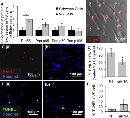
Figure 3.

NF‐κB is aberrantly activated in primary VS cultures and its siRNA‐mediated knockdown decreases proliferation. A. NF‐κB expression in cultured human VSs (n ≥ 6 tumors) normalized to expression in SC cultures (n ≥ 6 nerves) as quantified through western blot analysis. P‐means phosphorylated protein. Error bars represent SD. B. Representative image of effective transfection of a fluorescently labeled oligonucleotide (oligo, red) in primary VS cells. C. Representative proliferation images are shown for (a) scrambled siRNA or (b) siRNA treated primary VS cells. BrdU in nuclei (red) marks proliferating cells. D. Quantification of proliferation changes after siRNA treatment in primary VS cells normalized to proliferation in control scrambled siRNA treated (NT) cells (n = 4 results were from 4 independent results from cultures of two different patients). E. Representative cell death images are shown for (a) scrambled siRNA and (b) siRNA treated primary VS cells. TUNEL (green) in nuclei marks dying cells. F. Quantification of cell death rate after siRNA treatment of primary VS cells as measured by TUNEL staining (n = 3 different cultures). Error bars represent SD for panels D and F. *p = 0.025, re = compared to. Nuclei are labeled with Hoechst (blue) in (C, E).
