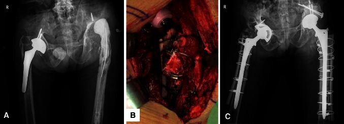
Fig. 1A–C.
A 45-year-old man had Paprosky Type IV bone defects of both hips. (A) His AP radiograph shows a loose porous-coated anatomic cementless femoral component and dislocated cement spacer of the left hip before revision surgery. (B) An intraoperative photographs shows the allografts are fixed with four Dall-MilesTM (Stryker Orthopaedics, Mahwah, NJ, USA) cables. (C) An AP radiograph of both hips taken 2 weeks after revision surgery shows the femoral and acetabular components are well fixed in a satisfactory position. The allografts are attached to the host bone with Dall-MilesTM cables.

