Figure 2.
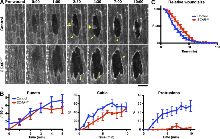
SCAR function in actin remodeling at wound edges. (A) Control and SCARΔ37 zygotic mutant embryos expressing GFP-Moesin were wounded and subjected to time-lapse imaging. Time points after wounding (minutes and seconds) are indicated. Note that although the actin puncta (arrowheads) and cable (single allows) appear in both the control and mutant embryos, the formation of actin protrusions (double allows) is severely reduced in the mutant, making its wound circumference markedly smoother than that of control. See also Video 2. Bar, 10 µm. (B) Quantitation of wound edge actin puncta, cable, and protrusion levels in control and zygotic SCARΔ37 embryos in the early phase of wound closure. n = 7–9 embryos. (C) Quantitation of wound closure in control and SCARΔ37 zygotic mutant embryos. Wound areas were normalized to the value at 5 min after wounding and plotted against time, as in Fig. 1 F. n = 4–17 embryos. Graphs show means ± SEM of the data.

