Figure 8.
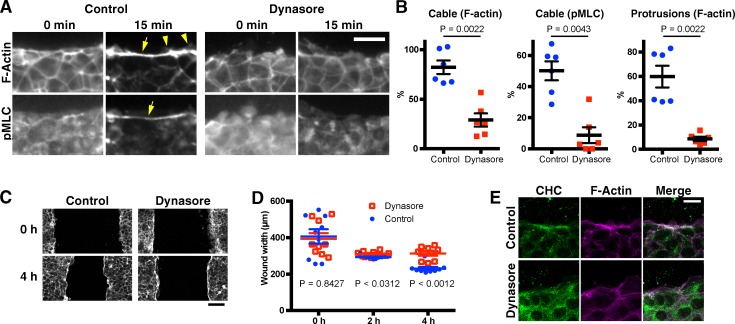
Dynasore inhibits wound edge actin remodeling by mammalian cells. Confluent monolayers of mIMCD3 mouse kidney epithelial cells were scratch wounded after treatment with DMSO (control) or 80 µM dynasore, a chemical inhibitor of Dynamin. At the indicated time points, the cells were fixed and stained with rhodamine-phalloidin and antibodies against phospho (=activated) myosin light chain (pMLC) or clathrin heavy chain (CHC), to examine the formation of an actomyosin cable and actin protrusions at the wound edge and the intracellular localization of clathrin. (A) Representative phalloidin and anti-pMLC images of control and dynasore-treated cells at 0 and 15 min after wounding. Arrows and arrowheads indicate the actin (myosin) cable and protrusions, respectively. (B) Cable and protrusion formation in the images 15 min after wounding was quantified. Cable formation was quantified using both the F-actin and pMLC images. Protrusions were quantified using the F-actin images. Note that the formation of both actin cable and protrusions are inhibited by dynasore. (C) Low magnification phalloidin images at 0 and 4 h after wounding. Note that the advancement of the cell sheets is inhibited by dynasore. At 4 h, a wound edge actin cable is observed, even for dynasore-treated cell sheets. (D) Quantification of wound width in each sample at the indicated time points. (E) Representative phalloidin and anti–clathrin heavy chain images of control and dynasore-treated cells at 15 min after wounding. Bars in the column scatter plots indicate means ± SEM of all plotted values. Line graphs show means ± SEM of the data. Bars: (A and E) 20 µm; (C) 100 µm.

