Figure 7.
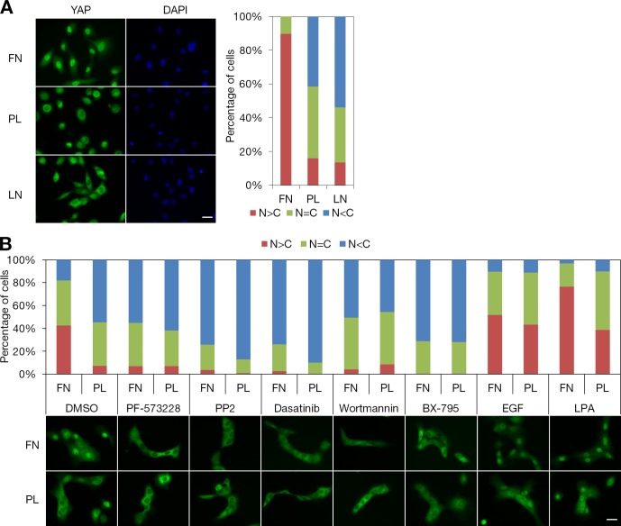
Attachment to fibronectin-coated coverslips induces YAP nuclear accumulation via the FAK–Src–PI3K–PDK1 pathway. (A) Attachment to fibronectin, poly-d-lysine, or laminin. Serum-starved MCF-10A cells were dissociated with Accutase and sparsely seeded on fibronectin (FN)-, poly-d-lysine (PL)–, or laminin (LN)-coated coverslips in starvation medium. Cells were incubated for 2 h before fixation. Subcellular localization of YAP was identified by immunofluorescence staining and quantified based on the criteria shown in Fig. 1 E. More than 170 cells from eight random views were quantified. DAPI staining was used to locate the nucleus. We performed three independent experiments. (B) Effects of inhibitors and growth factors on attachment-induced YAP nuclear localization. Serum-starved MCF-10A cells were detached and seeded on fibronectin- or poly-d-lysine–coated coverslips in starvation medium. After 3 h of incubation, cells were treated with the indicated inhibitors or mitogens for 30 min. Subcellular localization of YAP was identified by immunofluorescence staining. More than 120 cells from eight random views were quantified and confirmed in three independent experiments. Bars, 25 µm.

