Figure 8.
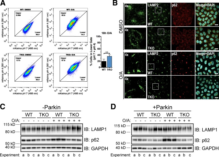
Concurrent deletion of TFEB, MITF, and TFE3 results in lysosomal and mitophagy defects. (A, left) WT and TFEB/MITF/TFE3 TKO (TKO) cells stably expressing YFP-Parkin and mt-mKeima were treated with DMSO or O/A for 18 h and subjected to FACS analysis. Plots are representative of n = 4 experiments. (A, right) Proportion of cells in the lower gate in left panels after 18-h O/A treatment. Data are means ± SD. **, P < 0.01. (B) WT and TKO cells stably expressing mCherry-Parkin were treated with DMSO or O/A for 18 h, fixed, immunostained for p62 and LAMP2, and analyzed by immunofluorescence. Left-most panels are enlarged versions of boxed regions in LAMP2 panels. Images are representative of n = 3 experiments where z stacks of five fields per condition (40–50 cells/field) per experiment were collected. Bars, 10 µm. (C and D) WT and TKO cells lacking endogenous Parkin (C) or stably expressing mCherry-Parkin (D) were treated with DMSO or O/A for 18 h, lysed, and immunoblotted. Three independent experiments (a–c) are shown.

