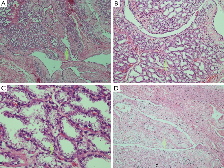
Figure 3.
Histologic features of the resected specimen. (A,B) The proliferation of lobules separated by hypercellular stroma and epithelial proliferation in cleft-like spaces (40×); (C) the lobules were lined by vacuolated secretory cells and contained eosinophilic secretions in the lumens (400×); (D) area of hemorrhagic necrosis within the tumor (100×).

