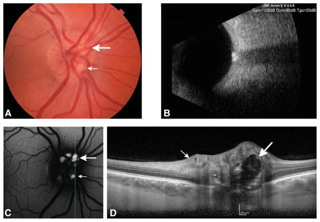
FIG. 2.
Fundus photograph (A), B-scan ultrasound (B), fundus autofluorescence images (C) and enhanced depth imaging optical coherence tomography (EDI-OCT) (D) of the left eye of subject with optic nerve head drusen. The green line in the red-free image indicates the direction of the EDI-OCT line scan. Superficial (small arrows) and buried drusen (large arrows) are shown.

