FIG. 3.

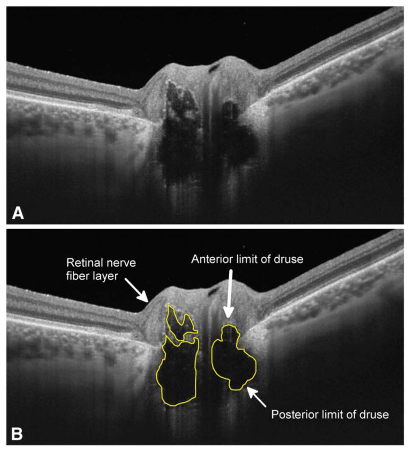
Enhanced depth imaging optical coherence tomography images of left eye of a subject with optic nerve head drusen (A), with the borders of the drusen outlined in yellow (B).

Enhanced depth imaging optical coherence tomography images of left eye of a subject with optic nerve head drusen (A), with the borders of the drusen outlined in yellow (B).