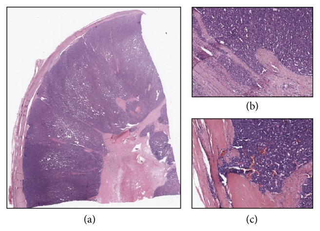
Figure 2.

Representative images of a follicular carcinoma previously treated with RFA. (a) The lower magnification (0.8x) picture shows scattered areas of hyaline sclerosis and scarring due to RFA, which do not affect the capsule. (b-c) The higher magnification (4x and 10x, resp.) pictures show spots of capsular invasion.
