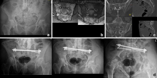
Fig. 2.

FFP type IIa. 84-year-old female with immobilizing lower back pain. Conventional radiograph did not show a bony lesion (a). Also with adequate pain medication mobilization was not possible. The MRI (T1 and STIR sequence in the coronal plane of the sacrum) showed bilateral bone bruise in the sacral ala with a transverse connection on level S2/S3 (b). A CT scan confirmed bilateral sacral involvement without fracture of the anterior pelvic ring (c). The patient was stabilized percutaneously with a trans-sacral bar and bilateral SI-screws (d)
