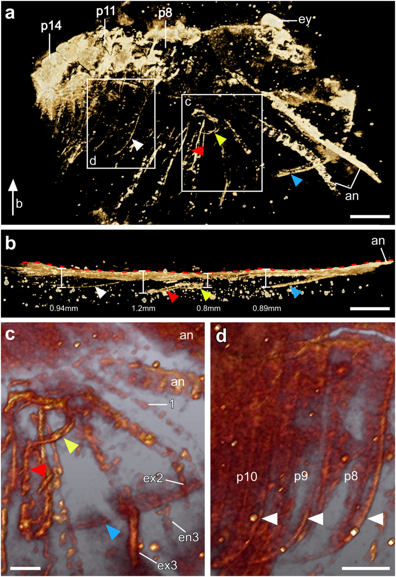
Figure 3. Fine details revealed inside the slab.
(a) Overview of the main part of the specimen. Micro-CT (Hires mode, i.e. high-resolution mode, Drishti) reveals the lateral margin (blue arrowhead) of the head shield. The annulation of the antennae is more clearly shown here than with light or fluorescence microscopy (cf. Fig. 1). The pleura of several consecutive segments (white arrowhead), and an opening (yellow arrowhead) and a fissure (red arrowhead) in the head shield were revealed. The white arrow indicates the angle from b. (b) 90°-rotated anterior part of the specimen shown in a (Hires mode, Drishti). The red dashed line indicates the surface of the specimen. The greatest depth of the lateral margin of the head shield (blue arrowhead) is 0.89 mm, that of the opening (yellow arrowhead) is 0.8 mm, the fissure (red arrowhead) 1.2 mm, and the pleurae (white arrowhead) 0.94 mm. (c) Close-up (volume rendering, Amira) of one side of the head shield from a showing an opening (yellow arrowhead) with a fissure (red arrowhead) extending towards the lateral margin (blue arrowhead) of the head shield; (d) Close-up (volume rendering, Amira) of the eighth to tenth post-antennal segments (p8–p10) indicated by their respective pleura (white arrowheads). Abbreviations as in Fig. 1. Scale bars, 2 mm. Photographs in in a, b taken by Y.L. and in c, d taken by G.S.

