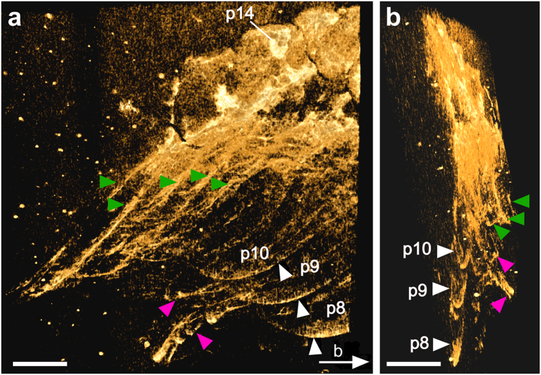
Figure 4. Fine details revealed in the posterior part of the specimen.
(a) Overview of the posterior part of the specimen. Micro-CT (Hires mode, Drishti) reveals pleurae from the right (green arrowheads) and left (white arrowheads; p8–p10) sides of the body. Two appendages (magenta arrowheads) are also visible. The white arrow indicates the viewing angle from b. (b) Posterior view of the posterior part of the specimen shown in a. The two appendages (magenta arrowheads) are located between the right and left (p8–p10) pleurae. While the right pleurae were preserved on the surface of the slab, the appendages (magenta arrowheads) and the left pleurae of the 9th to 11th post-antennal segments (p8–p10) were inside the slab. Abbreviations as in Fig. 1. Scale bars, 2 mm. Photographs in a, b taken by Y.L.

