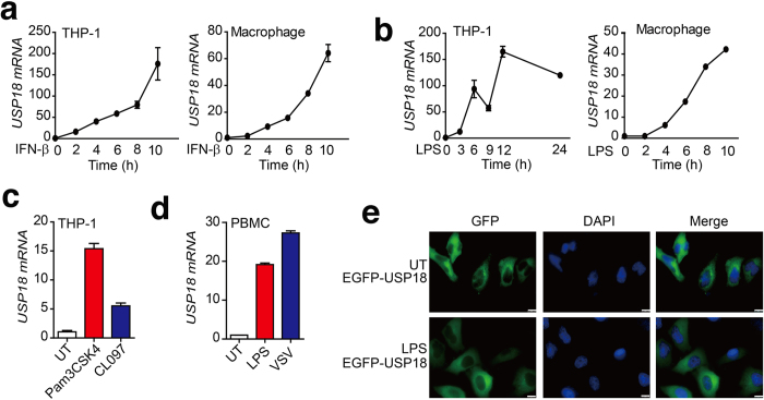
Figure 7. Expression and intracellular localization of human USP18 in response to TLR ligands stimulation.
(a-b) Relative USP18 mRNA abundance in THP-1 and THP-1-derived macrophage cells were determined by real-time PCR analysis after treatment with IFN-β (10 ng/ml) (a) or LPS (200 ng/ml) (b) for the indicated time points. (c) THP-1 cells stimulated by Pam3CSK4 (200 ng/ml) or CL097 (1 μg/ml). USP18 mRNA abundance was determined by real-time PCR analysis. (d) Human peripheral blood mononuclear cells (PBMCs) were treated with LPS or infected with VSV, and USP18-mRNA was determined by real-time PCR analysis. (e) Subcellular localization of human USP18 in HeLa cells, with or without LPS treatment. HeLa cells transfected with EGFP-USP18 plasmid were counterstained with DAPI and photographed under a fluorescence microscope. UT, untreated. Scale bar: 10 μm.

