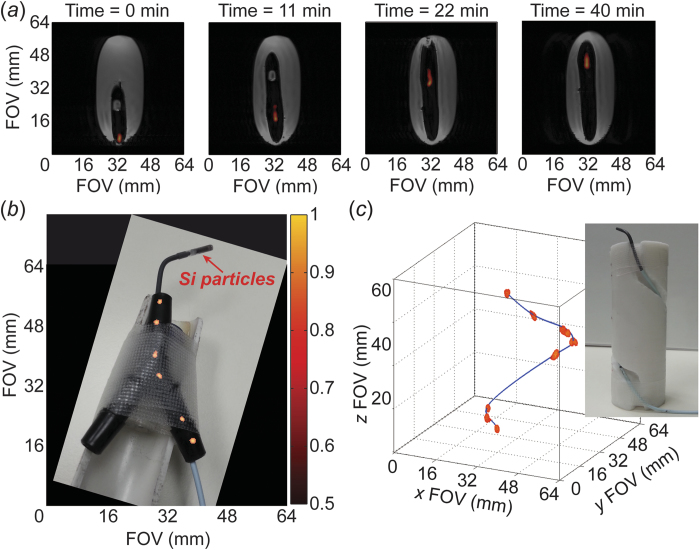
Figure 1. HP 29Si particle MRI-tracking in phantoms.
(a) Transit of ~50 mg of silicon particles loaded into a 24 Fr urinary catheter moving ~4 cm through a gelatin phantom over the course of 40 minutes; co-registered 29Si/1H imaging shows the outline of the catheter in the void space left in the gelatin. (b) Angiocatheter (5 Fr) loaded with ~6 mg of silicon particles moving through Y-shaped hollow plastic phantom to simulate branching of vasculature; picture of catheter and phantom superimposed with a composite of 29Si MRI images. The sample tube containing silicon particles is push-fit onto the tip of the angiocatheter. (c) Angiocatheter tracking three-dimensional passage around a spiral phantom (picture inset). Absolute 29Si signal intensities (colored scale, arbitrary units) are consistent for (a)–(c); greyscale denotes 1H intensities. Pertinent imaging parameters, as well as Supplemental Video S1 (showing a rotating view of Fig. 1c), are included in the Supplementary Materials.

