
Fig. 2.
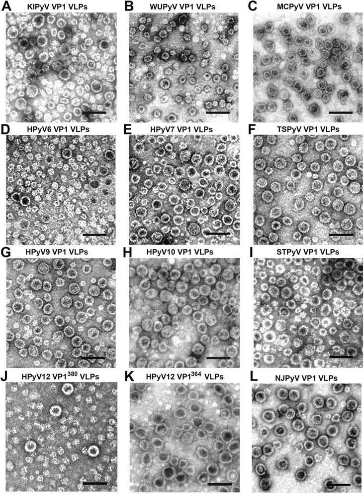
Detection of purified HPyV-derived VP1 VLPs using negative staining electron microscopy. Electron micrographs of: (a) - KIPyV VP1 VLPs, (b) - WUPyV VP1 VLPs, (c) - MCPyV VP1 VLPs, (d) - HPyV6 VP1 VLPs, (e) - HPyV7 VP1 VLPs, (f) - TSPyV VP1 VLPs, (g) - HPyV9 VP1 VLPs, (h) - HPyV10 VP1 VLPs, (i) - STLPyV VP1 VLPs, (j) - HPyV12 VP1380 VLPs, (k) - HPyV12 VP1364 VLPs, and (l) - NJPyV VP1 VLPs produced in yeast. Bars, 100 nm