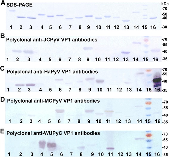
Fig. 5.

Detection of purified HPyV-derived VP1 VLPs in Western blot with polyclonal antibodies. a, Coomassie blue-stained SDS-PAGE; (b, c, d, e), Western blot with anti-JCPyV VP1 (b), anti-HaPyV VP1 (c), anti-MCPyV VP1 (d), and anti-WUPyV VP1 (e) polyclonal antibodies. The same samples of purified proteins were run on each gel. In lanes: 1 - HPyV16 L1 protein; 2 - BKPyV VP1 protein; 3 - JCPyV VP1 protein; 4 - KIPyV VP1 protein; 5 - WUPyV VP1 protein; 6 - MCPyV VP1 protein; 7 - HPyV6 VP1 protein; 8 - HPyV7 VP1 protein; 9 - TSPyV VP1 protein; 10 - HPyV9 VP1 protein; 11 - HPyV10 VP1 protein; 12 - STLPyV VP1 protein; 13 - HPyV12 VP1 protein; 14 - NJPyV VP1 protein; 15. Protein weight marker (Thermo Fisher Scientific Baltics); 16 - HaPyV VP1 protein
