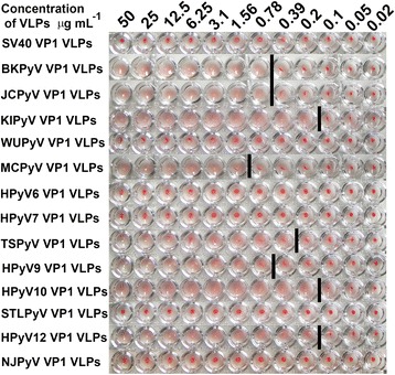
Fig. 6.

HA activity of HPyV-derived VP1 VLPs. A 2-fold dilution series of purified KIPyV-, WUPyV-, MCPyV-, HPyV6-, HPyV7-, TSPyV-, HPyV9-, HPyV10-, STLPyV-, HPyV12-, and NJPyV-derived VP1 VLPs expressed in S. cerevisiae were subjected to a HA assay with 1 % guinea pig erythrocytes. Purified JCPyV- and BKPyV-derived VP1 VLPs were used as positive controls, and SV40-derived VP1 VLPs were used as negative controls. The concentrations of VP1-derived VLPs in μg mL−1 are shown on the top. The bar indicates the highest VLP concentration at which HA of guinea pig erythrocytes was observed (HA titer)
