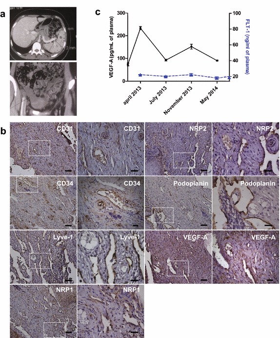
Figure 1.

a Radiological images of spleen (upper image) and pelvic region (lower image) at diagnosis. Multiple hypodense nodules are present in the spleen and fluid accumulation in the pelvic region is seen. b Immunohistochemistry of cervix tissue for blood and lymphatic vessel markers. Blood vessels were revealed by cluster of differentiation 31 and 34 (CD31, CD34), neuropilin-1 (NRP1, arterial marker), neuropilin-2 (NRP2, venous marker) immunostaining. Lymphatics were detected by Lymphatic vessel endothelial hyaluronan receptor-1 (LYVE-1) and Podoplanin immunostaining. Blood and lymphatic vessels are detected both by CD31 and NRP2 immunostaining. Furthermore, VEGF-A was significantly expressed in the tissue. Areas in the dotted squares are shown at higher magnification in the right hand images for each marker. Scale bars: Left panels 50 µm, right panels 20 µm). c Detection of vascular endothelial growth factor-A (VEGF-A) and soluble fms-like tyrosine kinase (FLT-1) in plasma samples by ELISA. VEGF-A (black dots) and FLT-1 (blue triangle). We noticed a decrease in VEGF-A levels between July 2013 and May 2014 (after Propranolol treatment) compared to April 2013 (before treatment). No significant change was observed for FLT-1. Vascular endothelial growth factor-C (VEGF-C) was below detection limit in all samples. Control levels of healthy donors were 75.78 pg/ml and 20.58 ng/ml for VEGF-A and FLT-1 respectively (pooled sample, n = 6).
