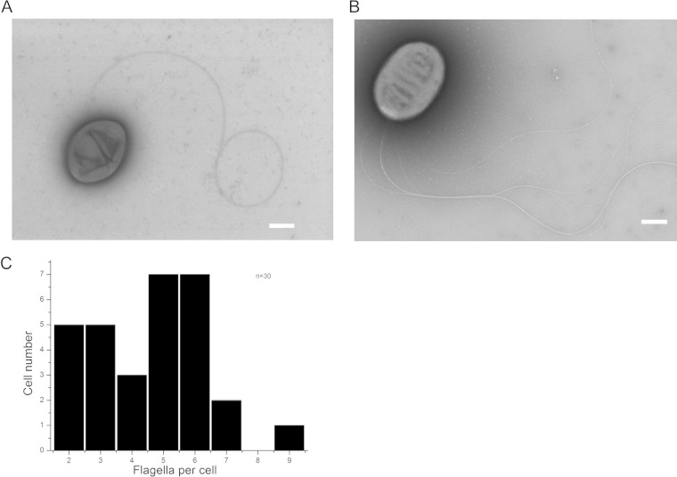
FIG 1.
Electron micrographs of R. sphaeroides strains WS8N and AM1. (A) Wild-type WS8N R. sphaeroides showing a medially located flagellum. (B) AM1 strain of R. sphaeroides displaying a bundle of polar flagella. Scale bars, 500 nm. (C) Graph showing the number of flagella displayed by AM1 cells. A total of 30 cells were scrutinized.

