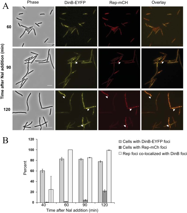
FIG 4.
Colocalization of Rep with Pol IV after Nal-induced DNA damage. (A) Phase-contrast, fluorescence, and overlay images of DinB-12L-EYFP (yellow) and Rep-mCh (red) focus formation and colocalization with time after Nal exposure. Scale bar = 5 μm. Arrows indicate representative colocalized foci. (B) Percentages of cells containing DinB-EYFP or Rep-mCh foci and percentages of Rep-mCh foci colocalizing with DinB-EYFP foci with time after Nal exposure. Shown are means ± SEM based on ∼200 cells (range = 186 to 274) counted at each time point in two independent experiments. Strain FC40 carrying plasmids pPFB913 and pPFB914 was grown in LB broth plus antibiotics at 37°C until mid-exponential phase (OD600 = 0.5), treated with 40 μg/ml Nal, and incubated for a further 2 h. Samples were withdrawn at 30-min intervals and visualized.

