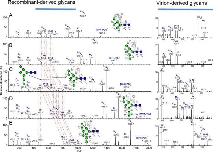
FIG 2.
Oligomannose isomers are the same on PBMC-derived Env and recombinant gp120. Shown are negative-ion CID spectra of oligomannose glycans from the recombinant gp120JR-CSF. The panels correspond to Man5GlcNAc2 (A), Man6GlcNAc2 (B), a mixture of Man7GlcNAc2 isomers (C), Man8GlcNAc2 (D), and Man9GlcNAc2 (E). The red dashed lines connect the fragment ions derived from the antenna linked to the 6-position of the core branching mannose residue (termed the 6-antenna), showing their shifts with increasing mannose residues in the 6-antenna of the larger glycans. The images on the right show spectra of the diagnostic region (m/z 450 to 1,000) from the virion-associated gp120 derived from PBMCs, confirming similarity in the glycans within the two samples.

