
FIG 4.
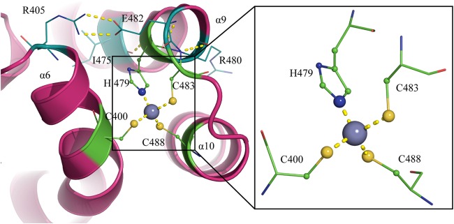
Zinc-binding motif of ICP27-CTD. A diagram representation of the zinc-binding motif shows the protein in magenta and the zinc ion as a gray sphere. The zinc ion is tetrahedrally coordinated by C400, H479, C483, and C488, shown by yellow dashed lines. H479, C483, and C488 form a helix-loop-helix structure. H479 is back-coordinated by E482, which in turn is stabilized by I475 and R405. R405 is in close proximity to the zinc-binding residue C400, providing more stability between helix and the helix-loop-helix. C483 is coordinated by R480.