FIG 2.
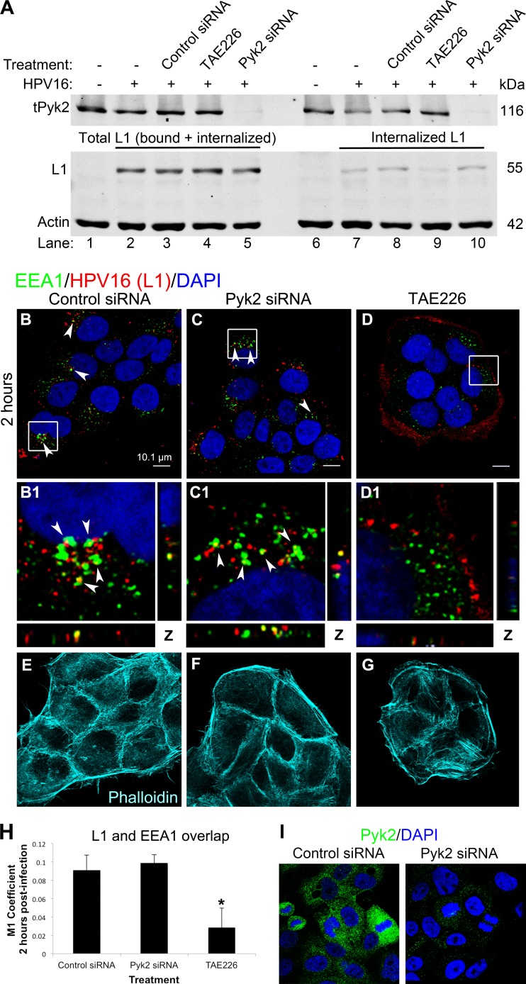
HPV16 internalization and early trafficking events are not disrupted in Pyk2-depleted HaCaT cells. (A) Western blot analysis of Pyk2, L1, and actin levels in HaCaT cells transfected with control or Pyk2 siRNA (lanes 3, 5, 8, and 10) or treated with 2 μM TAE226 and infected for 40 min with HPV16 PsVs. Cells were harvested directly for lysates (lanes 1 to 5) or treated with trypsin prior to harvesting (lanes 6 to 10). (B to G) Immunofluorescence analysis of EEA1 (green), L1 (red), and phalloidin (cyan) in cells transfected with control or Pyk2 siRNA or treated with 2 μM TAE226. Nuclei are stained with DAPI (blue). Colocalization of EEAI and L1 appears yellow. (H) The JACoP plugin for ImageJ was used to measure the M1 coefficient (fraction of red overlapping green) with three confocal scans for each condition. *, P < 0.05 (paired one-tailed t test). (I) Confirmation of siRNA-mediated Pyk2 knockdown via immunofluorescence analysis. HaCaT cells were transfected for 72 h with control or Pyk2 siRNA. Immunofluorescence analysis was performed with anti-Pyk2 antibody (green), and nuclei were stained with DAPI (blue).

