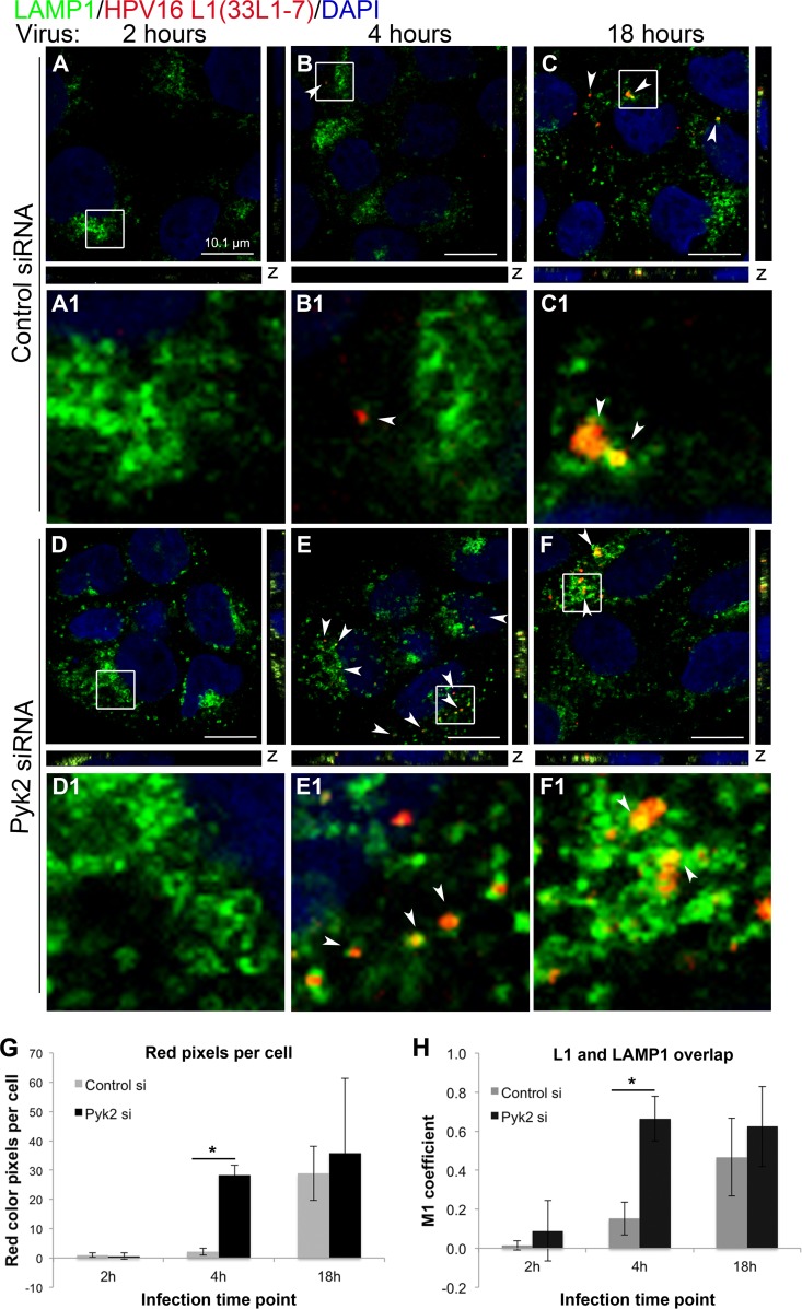
FIG 4.
Depletion of Pyk2 results in early unfolding of the HPV16 capsid in HaCaT cells. (A to C) Cells transfected with control nontargeting siRNA. (D to F) Cells transfected with Pyk2 siRNA. Cells were fixed at 2, 4, and 18 h postinfection with HPV16 PsVs. Immunofluorescence analysis was performed with anti-L1 33L1-7 (red) and anti-LAMP1 (green) antibodies. Nuclei were stained with DAPI (blue). Colocalization of L1 and LAMP1 appears yellow. (G) The color pixel counter plugin for ImageJ was used to measure red pixels, and the results were normalized to the number of cells in each scan. (H) The JACoP plugin for ImageJ was used to measure the M1 coefficient (fraction of red overlapping green) with three confocal scans for each condition. *, P < 0.05 (paired one-tailed t test).

