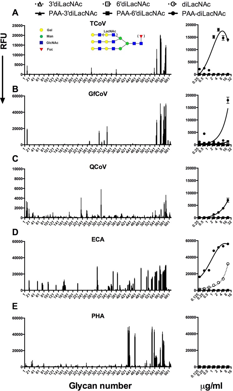
FIG 5.
Glycan binding specificity of avian coronavirus spike proteins. Shown are glycan binding specificities of S1 proteins of TCoV (A), GfCoV (B), QCoV d (C) and the plant lectins Erythrina cristagalli lectin (ECA) (D) and Phaseolus vulgaris agglutinin (PHA) (E). A representative example of the bound glycans for TCoV is depicted schematically. The left panels show the results of the glycan array; bar graphs were plotted in Prism and represent the averaged mean signal minus the background for each glycan sample, and error bars are the SEM values (from six replicates). The right panel shows the results of an ELISA-like assay in which the affinity of binding to sialylated and nonsialylated diLacNAc printed with and without conjugation to PAA was measured. RFU, relative fluorescence units.

