FIG 1.
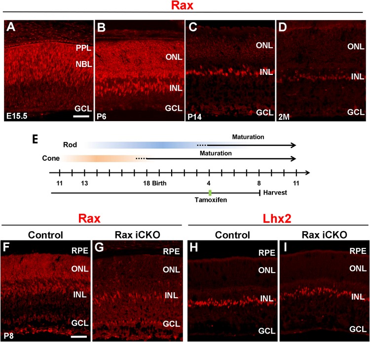
Expression and conditional inactivation of Rax in postnatal mouse retinas. (A to D) Mouse retinal sections were immunostained with an anti-Rax antibody (red) at E15.5 (A), P6 (B), P14 (C), and age 2 months (2M) (D). (E) Schematic diagram of schedule for tamoxifen administration and harvest of retinas. To conditionally inactivate Rax in postnatal photoreceptors, we treated Raxflox/flox; Crx-CreERT2 mice with tamoxifen at P4 and harvested the retinas at P8. (F to I) Retinal sections from Rax iCKO (P4 → P8) and control mice were immunostained with antibodies against Rax (red) (F and G) and Lhx2 (a Müller glia marker, red) (H and I). GCL, ganglion cell layer; INL, inner nuclear layer; ONL, outer nuclear layer; NBL, neuroblastic layer; PPL, presumptive photoreceptor layer; RPE, retinal pigment epithelium. Bars, 50 μm.

