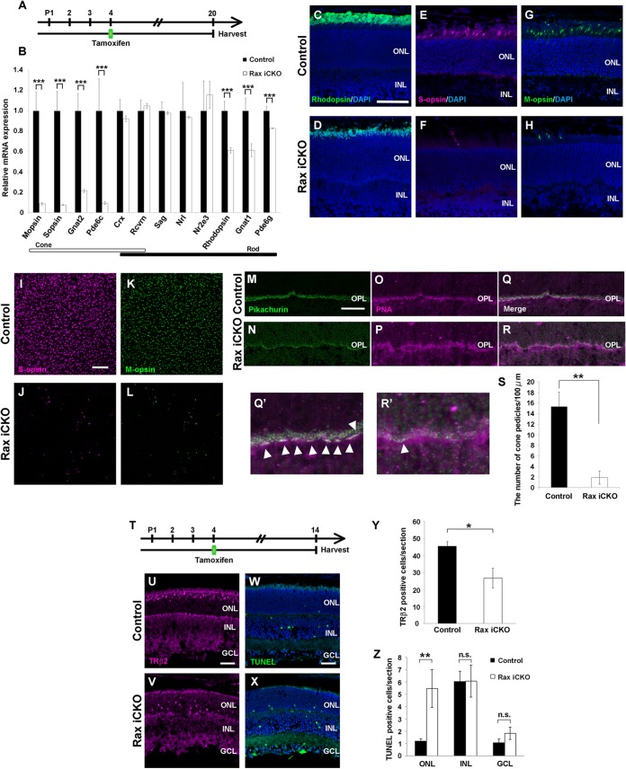
FIG 3.
Rax is required for cone photoreceptor cell survival during maturation. (A) Schematic diagram of schedule for tamoxifen administration and harvest of retinas. We treated Raxflox/flox; Crx-CreERT2 mice with tamoxifen at P4 and harvested the retinas at P20. (B) The expression levels of photoreceptor genes in Rax iCKO (P4 → P20) mouse retinas were analyzed by qPCR. The expression level of each was normalized to the expression level of a housekeeping gene, Gapdh (glyceraldehyde-3-phosphate dehydrogenase). The mean value for each control was set equal to 1.0. Error bars show ±SDs (n = 3). ***, P < 0.001. (C to H) Rax iCKO (P4 → P20) and control retinal sections were immunostained with the antibody against rhodopsin (green) (C and D), S-opsin (magenta) (E and F), or M-opsin (green) (G and H). The nuclei were stained with DAPI (blue). Bar, 50 μm. (I to S) Reduction of cone photoreceptor cells in the Rax iCKO (P4 → P20) mouse retina. (I to L) Whole-mount retinas from control and Rax iCKO (P4 → P20) mice were immunostained with the antibody against S-opsin (magenta) (I and J) or M-opsin (green) (K and L). (M and N) Retinal sections were immunostained with an antipikachurin antibody (a synaptic marker, green). (O and P) Cone pedicles were stained with PNA (magenta). (Q) Merge of panels M and O. (R) Merge of panels N and P. (Q′) and R′) Higher-magnification views of panels Q and R, respectively. Arrowheads, cone pedicles. Bars, 50 μm. (S) The number of cone pedicles decreased in the Rax iCKO (P4 → P20) mouse retina. Data are means ± SDs (n = 3). **, P < 0.01 by Student's t test. (T) Schematic diagram of schedule for tamoxifen administration and harvest of retinas. We treated Raxflox/flox; Crx-CreERT2 mice with tamoxifen at P4 and harvested their retinas at P14. (U to Z) Retinal sections from control (U) and Rax iCKO (P4 → P14) (V) mice were immunostained with an anti-TRβ2 antibody (guinea pig; magenta), a marker for developing cone photoreceptor cells. (W and X) TUNEL staining of retinas from control (W) and Rax iCKO (P4 → P14) (X) mice. Bars, 50 μm. (Y) The number of TRβ2-positive cells is shown. Data are means ± SDs (n = 3). *, P < 0.05. (Z) TUNEL-positive cells in control and Rax iCKO (P4 → P14) mouse retinas were counted. Data are means ± SDs (n = 3). **, P < 0.01 by Student's t test; n.s., not significant by Student's t test. INL, inner nuclear layer; ONL, outer nuclear layer; OPL, outer plexiform layer; GCL, ganglion cell layer.

