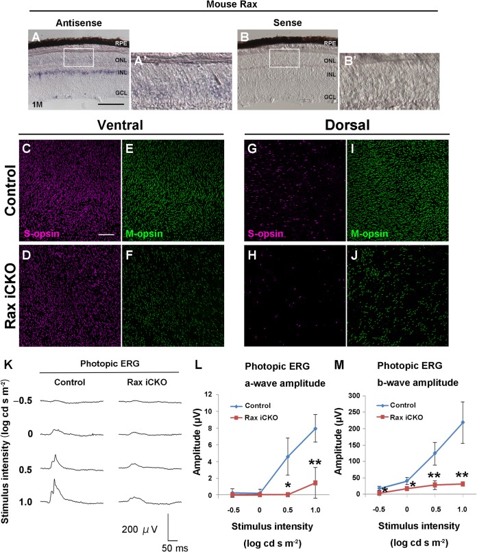
FIG 5.
Rax is required for maintenance of cone photoreceptors. (A to B′) Expression of Rax in the Raxflox/flox mouse retina at 1 month was detected by in situ hybridization using an antisense probe (A) or a sense probe (B) for Rax. (A′ and B′, magnifications of boxes in panels A and B, respectively. GCL, ganglion cell layer; INL, inner nuclear layer; ONL, outer nuclear layer; RPE, retinal pigment epithelium; 1M, harvest of retinas at age 1 month. Bar, 100 μm. (C to J) Whole-mount retinas from control and Rax iCKO (1 month → 2 months) mice were immunostained with antibody against S-opsin (magenta) (C, D, G, and H) or M-opsin (green) (E, F, I, and J). Ventral (C to F) and dorsal regions (G to J) are shown. Bar, 100 μm. (K to M) ERGs were recorded from Rax iCKO (1 month → 2 months) mice. (K) Photopic ERGs elicited by four different stimulus intensities are shown. (L and M) The amplitudes of the photopic ERG a-wave (L) and b-wave (M) are shown as a function of the stimulus intensity. For control mice, n = 5; for Rax iCKO mice, n = 3. Data are means ± SDs. **, P < 0.01 by Student's t test; *, P < 0.05 by Student's t test.

