FIG 11.
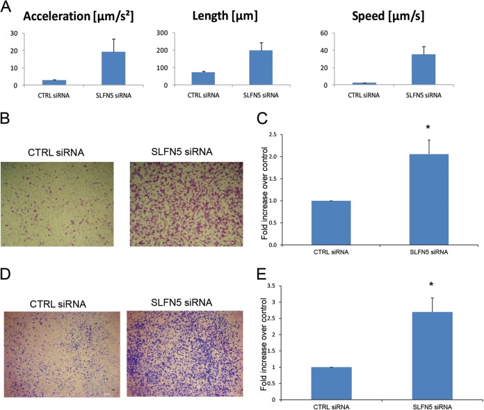
SLFN5 inhibits malignant cell migration. (A) 786-0 cells were transiently transfected with control siRNA or SLFN5 siRNA, as indicated; at 48 h after transfection, cells were transferred to a Nikon Biostation incubator wide-field microscope and time-lapse images were acquired at 10-min intervals for 12 h. Data were analyzed using the Nikon Elements software. Data represent the means ± standard errors of results from 27 migratory events detected by the Nikon Elements software. (B) 786-0 cells were transiently transfected with control siRNA or SLFN5 siRNA, as indicated. At 48 h after transfection cells, were plated in BD BioCoat cell culture inserts for 3 h and then cells were stained. Representative images of migrating cells (×5) after 3 h of incubation are shown. (C) The migrated cells were counted, and relative migration is expressed as the fold increase over control ± standard error of results from three independent experiments. Paired two-tailed t test analysis showed a P value of 0.0198 (*). (D) 786-0 cells were transiently transfected with control siRNA or SLFN5 siRNA. At 48 h after transfection, the cells were plated in a Corning BioCoat Matrigel invasion chamber, and invasion of the cells was assessed in 6 h. Representative images of invading cells (×5) after 6 h of incubation are shown. (E) The invading cells were counted, and relative invasion is expressed as the fold increase over controls ± standard errors of results from four independent experiments. Paired two-tailed t test analysis showed a P value of 0.02933 (*).

