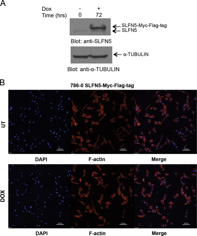
FIG 6.
Effect of SLFN5 overexpression on cell morphology. (A) 786-0 pLVX/tetONE-puro-SLFN5-Myc-Flag tag-transduced cells were treated with doxycycline (Dox) as indicated, and after cell lysis, proteins were resolved by SDS-PAGE and immunoblotted with an anti-SLFN5 antibody and an antitubulin antibody. (B) The same cells as used in panel A were treated with doxycycline for 72 h and then seeded onto 0.2% gelatin-coated glass coverslips. Cells were then fixed, permeabilized, and stained with Alexa Fluor 568-labeled phalloidin and 4′,6-diamidino-2-phenylindole (DAPI) to detect cytoskeletal F-actin and nuclei by confocal fluorescence microscopy. Representative areas showing the F-actin structure and nuclei are shown. Bars, 100 μm. UT, untreated.

