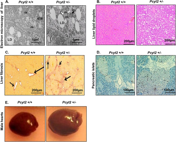
FIG 1.
Multiorgan abnormalities in the context of Pcyt2+/− metabolic syndrome. (A) Electron microscopy reveals an increased amount of liver lipid droplets (LD), reduced and damaged mitochondria (m) (black arrow), and mitophagy (m with two membrane layers) (white arrows) in 8-month-old Pcyt2+/− liver. (B and C) Liver sections stained with H&E (B) and Picrosirius red (C) reveal severe liver steatosis and fibrosis in Pcyt2+/− mice. Phenotypes in panels A to C are present in both male and female Pcyt2+/− mice. (D) Staining for somatostatin 14 and pancreatic polypeptide demonstrates pancreatic islet hypertrophy in Pcyt2+/− mice. (E) Representative images illustrating that 8-month-old Pcyt2+/− male hearts are larger than the hearts of control male littermates.

