FIG 2.
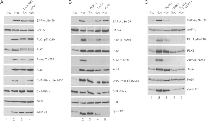
SAF-A S59 phosphorylation in nocodazole-treated cells requires PLK1 and/or CDK1. (A) Asynchronously growing HeLa cells were either left untreated (Asy) or treated with 40 ng/ml nocodazole for 16 h as for Fig. 1. One hour prior to mitotic shake-off, cells were untreated or treated with 8 μM DNA-PK inhibitor NU7441 or 5 μM ATM inhibitor KU55933. Cells were harvested by mitotic shake-off and left to recover for 35 min in fresh medium (without nocodazole) but in the presence of the protein kinase inhibitors as indicated. Cells were lysed using NETN lysis buffer containing 1% NP-40 with protease and protein phosphatase inhibitors. Fifty micrograms of whole-cell extract was run on an 8% gel and transferred to nitrocellulose, and blots were probed using the in-house-generated rabbit phospho-specific SAF-A S59 antibody and then with antibodies to SAF-A (Abcam number 10297 mouse), PLK1, PLK1-pT210, Aurora A and Aurora A-pT288, DNA-PKcs, DNA-PKcs-pS2056, Ku80 (loading control), and cyclin B1 as indicated. (B) Experiments were carried out as for panel A, but cells were incubated with 100 nM PLK1 inhibitor (BI2536), 100 nM Aurora A inhibitor 1 (AurA-i), or 100 nM Aurora B inhibitor (hesperadin) as indicated. (C) As for panels A and B, but cells were treated with the PLK1 inhibitor BI2536 at 100 nM (lane 3), the CDK1 inhibitor RO3366 at 20 μM (lane 4), or the PLK1 inhibitor BI2536 at 100 nM plus RO3366 at 20 μM (lane 5) as indicated.

