
FIG 3.
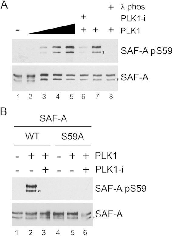
PLK1 phosphorylates SAF-A in vitro. (A) FLAG-tagged SAF-A WT was immunoprecipitated from asynchronously growing MRC5-CV cells as described in Materials and Methods and incubated under kinase assay conditions alone (lane 1) or with 0.1, 0.5, 1, or 2 μg of purified PLK1 (lanes 2 to 5, respectively). Lanes 6 to 8 contained 2 μg purified PLK1. In lane 6, the PLK1 inhibitor BI2536 was added to reaction mixtures (final concentration, 100 nM) prior to addition of PLK1. All reactions were stopped by addition of SDS sample buffer, and samples were run on SDS-PAGE and analyzed by Western blotting using the rabbit phospho-specific antibody to S59 and a rabbit polyclonal antibody to total SAF-A (Abcam number 20666). The band corresponding to full-length SAF-A runs at ∼125 kDa. The faster-migrating band, running at ∼110 kDa, indicated by the asterisk, likely represents either an alternatively spliced or a proteolytically cleaved form of SAF-A. In lanes 7 and 8, samples were incubated with lambda phosphatase buffer (lane 7) or lambda phosphatase (200 units) (lane 8) for 10 min prior to addition of SDS sample buffer and SDS-PAGE. (B) SAF-A WT or SAF-A S59A was immunoprecipitated from asynchronously growing MRC5-SV cells and incubated with purified PLK1 as described in Materials and Methods. Reactions were analyzed by SDS-PAGE followed by Western blotting and probed for SAF-A S59 phosphorylation and total SAF-A as indicated for panel A. The two bands cross-reacting with the SAF-A antibody are as in panel A.