
FIG 4.
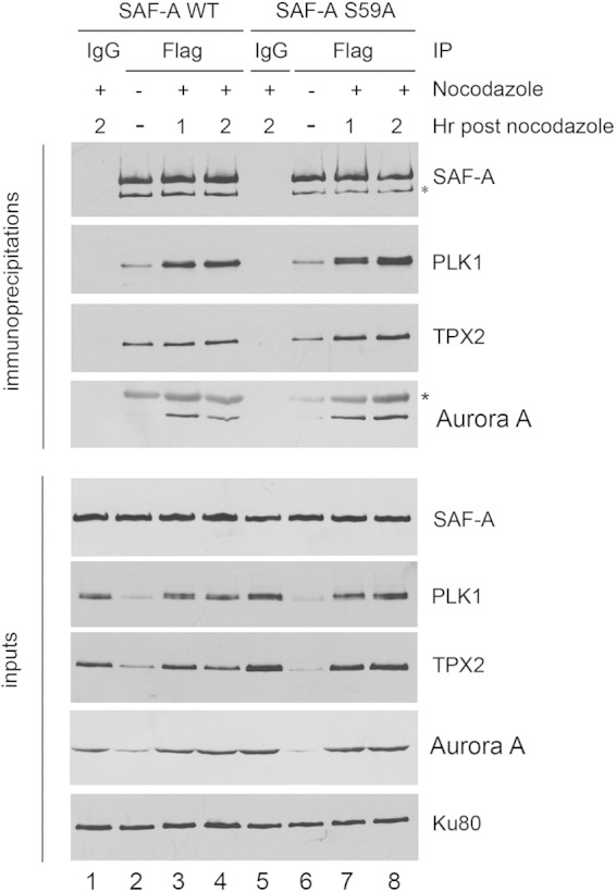
SAF-A interacts with PLK1, and the interaction does not require S59 phosphorylation. MRC5-SV cells expressing either FLAG-tagged SAF-A WT (lanes 1 to 4) or SAF-A S59A (lanes 5 to 8) were grown in the presence of nocodazole 40 ng/ml (16 h) (lanes 1, 3, 4, 5, 7, and 8) or under asynchronous conditions (lanes 2 and 6). The nocodazole was washed away, and cells were placed in nocodazole-free medium and harvested after either 1 h (lanes 3 and 7) or 2 h (lanes 1, 4, 5, and 8). SAF-A was immunoprecipitated using anti-FLAG beads, and samples were washed, run on SDS-PAGE, and analyzed by immunoblotting using the antibodies indicated. Samples in lanes 1 and 5 were from immunoprecipitates using an equivalent amount of IgG and protein G-Sepharose beads as a control. The lower panels show Western blots for 50 μg of protein for each immunoprecipitate. For immunoblotting of immunoprecipitates, a rabbit antibody to SAF-A was used. For inputs, a mouse monoclonal antibody to SAF-A was used. The two bands cross-reacting with the SAF-A antibody are as in Fig. 3A. The asterisk in the immunoblot for Aurora A indicates a cross-reacting band, likely immunoglobulin.