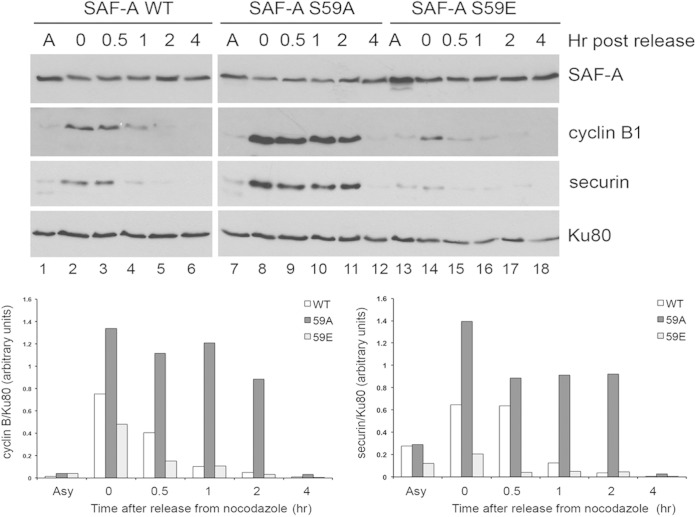
FIG 9.
Inability to phosphorylate SAF-A on S59 in nocodazole-treated cells results in enhanced and prolonged expression of cyclin B1 and securin. SAF-A WT, 59A, and 59E cells were grown to 70% confluence, and extracts were prepared from either asynchronously growing cells or cells treated with nocodazole (40 ng/ml for 16 h) and harvested by mitotic shake-off either immediately (0 h) or at various times after placement in fresh, nocodazole-free medium as indicated. Fifty micrograms of total protein from NETN extracts was run on SDS-polyacrylamide gels and transferred to nitrocellulose, and blots were probed with the indicated antibodies. The antibody to SAF-A was a mouse monoclonal antibody. Bands corresponding to cyclin B1 and securin were quantitated and normalized to total Ku80 and are graphically shown below gels on the left, and right, respectively. Results are representative of 3 separate experiments. See Fig. S10 in the supplemental material for similar results with the microtubule stabilizer paclitaxel.

