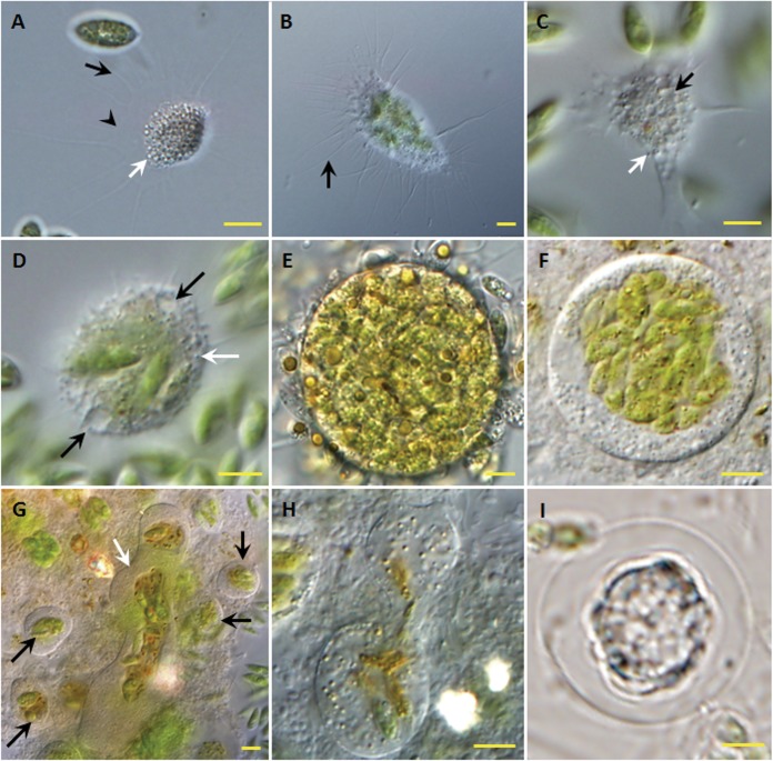
FIG 2.
Forms and morphological traits of V. algivore gen. nov., sp. nov. (DIC optics). (A) Advancing trophozoite with an expanded morphotype; long, thin, tapering, and mostly unbranched pseudopodia (black arrow) arise from a hyaline and very delicate fringe of the cytoplasm (arrowhead), and there are many inclusions (white arrow) inside the cell body. (B) Advancing trophozoite with ingested Scenedesmus cells; the pseudopodia (arrow) arise from a delicate fringe of cytoplasm. (C) Cell with numerous vacuoles (black arrow) and peripheral granules (white arrow). (D) Forming plasmodium with two contractile vacuoles (black arrows); small refractile granules (white arrow) lie just below the cell surface, and there is no thin hyaloplasm. (E) Large early digestive cyst with many Scenedesmus cells located throughout the cell. (F) Large digestive cyst in the middle stage, in which the ingested algal cells have moved to the center of the amoeba. (G) Digestive cysts with different shapes; most are small and round with centralized food (black arrows), and one large digestive cyst is elongate and irregular (white arrow) with a central mass of food. (H) Digestive cysts in division. (I) Resting stage. Scale bars = 5 μm.

