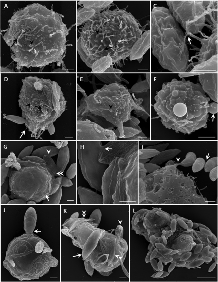
FIG 3.
Scanning electron micrographs of V. algivore. (A) Trophozoite, with many thin and long filopodia (arrow) and some retracted filopodia that became dome-shaped structures (arrowhead). (B) Larger trophozoite, with some filopodia (arrow) and more dome-shaped structures (arrowhead). (C) Detail of how filopodia capture prey; the arrow shows where the filopodium appears branched. (D) Trophozoite with shorter filopodia (white arrow) and some dome-shaped structures (arrowhead); one Scenedesmus cell (black arrow) is being engulfed. (E) Trophozoite with thick filopodia (arrow) and more dome-shaped structures (arrowhead). (F) Trophozoite with significant dome-shaped structures (arrow) on the surface of the cell. (G) Digestive cyst surrounded by an organic cyst wall (arrow), which was covered by Scenedesmus cells; in some, only an empty cell remained (double arrowheads). Algal cells can be connected to each other (arrowhead). (H) Detail of panel G showing that the cell walls were disrupted and fused (arrow). (I) Four Scenedesmus cells outside the amoeba were connected by unknown material (arrow), and one of them was connected by the cell inside the amoeba (arrowhead). This might be an artifact of preparation but is consistent with the information in Fig. 5H. (J) Round digestive cyst with thin membrane and one Scenedesmus cell adhering to the surface (arrow). (K) Large and irregular digestive cyst with some Scenedesmus cells, showing one cell (arrow) adhering to the surface, one shrunken cell submerged in the body (arrowhead), and several empty cells beneath the thin membranes (double arrowheads). (L) Large irregular digestive cyst with many adhering Scenedesmus cells. Scale bars = 2 μm (A to K) and 10 μm (L).

