FIG 4.
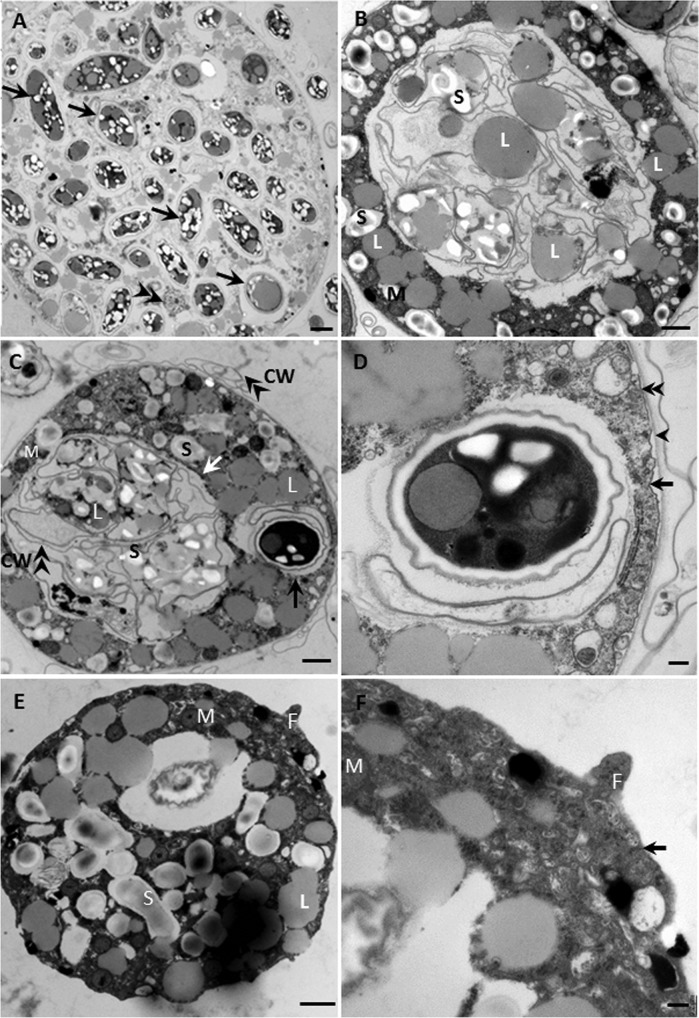
Transmission electron micrographs of thin-sectioned V. algivore. (A) Newly formed digestive cyst with many intact algal cells (arrows) inside the body and one nucleus (double arrowheads). (B) Middle-stage digestive cyst with one large food vacuole in the center of the body and lipids (L) and starch (S) distributed both inside and outside the food vacuole. (C) Late-stage digestive cyst with one large food vacuole (white arrow) and a newly formed food vacuole (black arrow); many algal cell walls (double arrowheads) lie inside the old vacuole, and some lie outside the cell. CW, cell wall; M, mitochondrion. (D) Detail of cell in panel C showing a digestive cyst with one intact Scenedemus cell with the cell membrane (arrow) and a cyst envelope (arrowhead); in some regions (double arrowheads), the cyst and the plasma membrane adhere to each other. (E) Trophozoite with many mitochondria, lipid, and starch inclusions. F, filopodium. (F) Detail of pseudopodium in panel E. There is a single membrane around the trophozoite, and the electron density of the filopodium is similar to that of the cytoplasm. Scale bars = 2 μm (A), 1 μm (B, C, and E), and 0.2 μm (D and F).

